toh

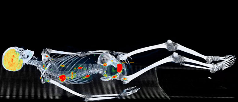
Nuclear Medicine and Molecular Imaging is a branch of medical imaging that uses radioactive products called tracers to check how well organs and tissues function within the body.
The radiation risk involved in these procedures is very low compared with the potential benefits. There are no known long-term adverse side effects from Nuclear Medicine and Molecular Imaging procedures, which have been performed for more than 50 years. Rarely, allergic reactions may occur but are extremely rare and usually mild.
In some cases, radioactive products can also be used to treat disease.
If you cannot make you appointment, it is important that you notify the department at 613-761-4831 as soon as possible. Failure to make your appointment results in wasting of expensive materials that are ordered especially for your appointment and also reduces availability to other patients. Missed appointments may also result in delays of your treatments.
How does it work?
For most procedures, you will receive a small amount of a radioactive “tracer” through a vein in your arm, by mouth or occasionally by breathing it in as a mist. The amount of radioactivity is very small, and it is unlikely that you will experience any side effects or discomfort.
When the radioactive tracer travels to the part of the body to be examined, pictures (scans) are obtained using a gamma camera. A gamma camera sees radioactivity in a similar way that an ordinary camera sees light. The procedure will feel no different from having your photograph taken but the exposure time will be longer.
While having the pictures taken you will need to sit or lie still on a special table. The camera may come close to you but will not touch you.
You will hear the noise of machinery moving the camera and other electronic sounds. It is like having an X-ray taken, but no radiation comes from the camera.
A technologist will be in the room with you for most of the time to operate the gamma camera, attend to your needs and answer your questions. They may leave the room briefly but will let you know when this is necessary.
Before you leave the department, the technologist will check the pictures to be sure that the test is complete and has provided the most useful results possible.
What is it used for?
Most often, radioactivity is used to show the presence and extent of a disease or condition. This information will help your doctor with diagnosis and treatment. Less often, radioactivity is used for treatment (e.g., thyroid disease, prostate disease).
We carry out Nuclear Medicine and Molecular Imaging tests and treatments only at the request of your doctor.
Radioactive materials have been used in this way for more than fifty years. Many conditions and diseases are investigated by Nuclear Medicine and Molecular Imaging techniques.
Very small quantities of radioactivity are used in diagnostic tests. The tests are like the familiar x-ray procedures and usually involve obtaining pictures (scans) of the affected part of the body.
There are different types of nuclear medicine images:
- There are different types of nuclear medicine images:
- Dynamic – a series of images that captures movement or activity (such as blood flow to an organ)
- Planar (static) – a 2-dimensional view that shows one image at a time
- Whole body – front and back views of the body in 2 dimensions
- Single photon emission computed tomography (SPECT) – a 3- dimensional view of the process or function of the organ being studied
- Positron Emission Tomography (PET) – a 3-dimensional view of the body
- CT – CT imaging may be used in some examinations for accurate localization
Nuclear Medicine and Molecular Imaging Clinical Uses
- It helps in early diagnosis.
- Determines severity of disease.
- Helps select the most effective therapy.
- Identifies recurrence of disease.
- Assesses and evaluates the progression of disease.
Different radioactive isotopes are used depending on the type of test and the tissue or organ being studied, including:
- Technetium (Tc-99m)
- Radioactive iodine (I-131 or I-123)
- Gallium (Ga-67 or Ga-68)
- Indium (In-111)
- Lutetium (Lu-177)
- Radium (Ra-223)
- Fluorine (F-18)
Generally, no special preparations are required, but for some studies it is necessary to go without breakfast or to stop taking certain medications. If special preparation is required in your case, you will receive instructions. If in doubt, telephone us at the location (campus) where your procedure is scheduled.
If you are or think that you might be pregnant, or if you are breastfeeding, please notify us. The procedure may have to be modified, postponed or cancelled.
You may save time by wearing loose, comfortable clothing such as a sweat suit. Please do not wear jewelry, hairpins, or metal belt buckles. If your schedule requires that you arrive in more formal attire, you may be asked to change into a hospital gown.
Many Nuclear Medicine procedures are divided into different phases separated by an hour or more (may be days) waiting period after the injection of a radioactive “tracer” before pictures (scans) can be obtained. You may want to bring something to read. In some cases, you may leave the department or the hospital.
Please remember to bring your provincial Health Card to your appointment.
Central Booking office for Nuclear Medicine and Molecular Imaging: (613) 761-4831*
* Press the appropriate “option” to connect with the imaging modality (i.e. CT, MRI etc).
| Civic Campus 1053 Carling Avenue – 1st Floor Tel.: 613-761-4280 Hours: Mon. – Fri., 8:00 a.m. – 4:00 p.m. Directions From within the Civic Campus, take the “C” elevators to the 1st Floor and follow the signs to Nuclear Medicine and Molecular Imaging. Patients may also ask for directions at the patient information desk. |
| General Campus 501 Smyth Road Main level Tel.: 613-737-8395 Hours: Mon. – Fri., 8:00 a.m. – 4:00 p.m. Directions From the main entrance, follow the signs on the main level (located at the public elevators). Patients may also ask for directions at the patient Information desk. |
What if I must cancel or reschedule my appointment?
If you cannot make you appointment, it is important that you notify the department at 613-761-4831 as soon as possible. Failure to make your appointment results in wasting of expensive materials that are ordered especially for your appointment and also reduces availability to other patients. Missed appointments may also result in delays of your treatments.
Should you be admitted to the hospital, please inform your doctor about your appointment.
Will I be OK to drive or go home on my own?
Because no reaction to the radioactive tracer is expected, you may be able to drive home if you wish. Alternatively, a relative or friend may accompany you. For some examinations, you may receive a drug for which driving afterward is prohibited. You will be informed in advance if this is the case for your examination.
Will there be any after-effects?
Nuclear Medicine and Molecular Imaging procedures are not difficult and are very safe. These procedures use small amounts of radioactive materials to image different parts of the body.
It is unlikely. Only a small dose of radiation is used. The dose is kept to the minimum necessary to obtain a useful result. The risk from this radiation is extremely small.
Many Canadians benefit each year from Nuclear Medicine and Molecular Imaging procedures used to diagnose and treat a wide variety of diseases and conditions.
How are radiopharmaceuticals use?
Nuclear Medicine and Molecular Imaging procedures require that a radiotracer be injected, swallowed, or inhaled. Each radiotracer is attracted to specific organs, bones, or tissues. A special camera (PET, SPECT, or gamma camera) takes pictures of the distribution of the radiotracer in the body.
The use of radiation in these procedures offers a safe means to provide doctors with diagnostic information that would otherwise require exploratory surgery or more expensive or difficult procedures to obtain the same information.
Radiopharmaceuticals are also used for therapy, to treat overactive thyroids and certain types of cancer.
Radiopharmaceuticals are approved by Health Canada and are tested carefully prior to general use and prepared with great care.
Tracers are often made especially for you and can be very costly. If you cannot make you appointment, it is important that you notify the department at 613-761-4831 as soon as possible.
How much radiation exposure is involved in Nuclear Medicine and Molecular Imaging procedures?
Because the amount of radiotracer used in Nuclear Medicine and Molecular Imaging tests are extremely small, the patient’s radiation exposure is small.
Nuclear Medicine and Molecular Imaging specialists and technologists use the ALARA principle (As Low As Reasonably Achievable) to carefully select the amount of radiopharmaceutical that will provide an accurate test with the least amount of radiation exposure to the patient. The actual dosage is determined by the reason for the study and the body part being imaged.
The amount of radiation in most Nuclear Medicine and Molecular Imaging tests is comparable to and often less than that of a diagnostic CT scan. Most Nuclear Medicine and Molecular Imaging procedures expose patients to about the same amount of radiation as they receive in a several months of normal living.
Who performs the exam?
A specially trained Nuclear Medicine and Molecular Imaging technologist will perform the procedure under the direction of the Nuclear Medicine and Molecular Imaging doctor.
How and when will I know the results?
After examining your test results, a Nuclear Medicine and Molecular Imaging physician will send a written report to your physician. Your physician will explain the results to you when you see her/him.
Tests and Procedures
- Adrenal Medulla MIBG Scan
- Bone Mineral Density (BMD) Scan
- Bone Marrow Scan
- Bone Scan
- Brain Scan
- Breast Lymphangiogram Study
- C-14 Urea Breath Test
- Cardiac Amyloidosis Scan
- Cardiac Perfusion Scan (Day 1)
- Cardiac Perfusion Scan (Day 2)
- CSF Leak Study
- CSF Study (Radionuclide Cisternogram)
- DaTscan Study
- Esophageal Transit/Aspiration study
- Gallium scan
- Gastric Emptying Study (Liquid)
- Gastric Emptying Study (Solid)
- Gastrointestinal Bleeding (GI Bleed) Study
- Heat Damaged RBC Spleen Study
- Hepatic Hemangioma Study
- Hepatobiliary study (HIDA)
- Lacrimal Duct Scan (Dacryoscintigraphy)
- Liver/Spleen Scan
- Lung (Ventilation/Perfusion or V/Q) Scan
- Lymphangiogram Scan
- Meckel’s Diverticulum Scan
- Melanoma Lymphoscintigraphy Study
- MUGA: Gated Cardiac Scan
- Octreotide Scan
- Parathyroid Scan
- FDG PET/CT scan
- PSMA PET/CT Scan
- Quantitative Lung Scan
- Radioactive Iodine Ablative Therapy for Thyroid Cancer
- Radioactive Iodine (RAI) Treatment for Hyperthyroidism
- Renal Captopril Scan
- Renal Cortical Scan
- Renal GFR (Glomerular Filtration Rate) Study
- Renal Scan
- Right to Left Shunt Study
- Salivary Gland Scan
- SeHCAT Scan
- Shunt Patency Study
- Thyroid Scan and Uptake Study
- Vulvar Cancer Lymphoscintigraphy Study
- White Blood Cell Imaging (WBC scan)
- Whole–Body Iodine Scan
- Xofigo Radium-223 Therapy
Last updated on: March 15th, 2024


 To reset, hold the Ctrl key, then press 0.