toh

What is Ultrasound?
Diagnostic Ultrasound is an imaging procedure using high-frequency sound waves rather than x-rays to create pictures.
The sound wave frequency used is too high to be felt or heard. Many organs and body parts can be seen using ultrasound.
How Is It Done?
Most ultrasound examinations are done using a sonar device outside your body, though some ultrasound examinations involve placing a device inside your body.
Why is it Done?
Ultrasound is used to help doctors to diagnose illness or to guide procedures
- Diagnose the disease in the abdomen and pelvis
- Evaluate flow in blood vessels
- Guide a needle for biopsy or other procedure
- Evaluate a breast lump
- Evaluate the thyroid gland
- Evaluate musculo-skeletal structures
Safety Information
Diagnostic ultrasound is a safe procedure that uses sound waves to create the image. There are no known risks.
Although ultrasound is a valuable tool, it has limitations. Sound doesn’t travel well through air or bone, so ultrasound isn’t effective at imaging parts of your body that have gas in them or are hidden by bone.
To view these areas, your doctor may order other imaging tests, such as CT, MRI scans or X-rays.
Exam Information
What happens to me during the procedure?
During an ultrasound exam, you may need to remove the jewelry and some or all of your clothing, change into a gown, and lie on an examination table. The procedure will be performed in a darkened room.
When in the room, you will be asked to lie on a narrow table, and the sonographer will explain the procedure to you and may ask you pertinent medical questions. The gel is applied to your skin to establish good contact with your skin and eliminate air pockets.
What can I expect?
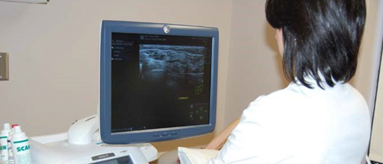
- A trained sonographer presses a small, hand-held device (transducer), about the size of a bar of soap, against your skin over the area being examined, moving it as necessary to capture the image.
- The transducer sends sound waves into your body, collects sound waves that bounce back and sends them to a computer, which creates the images.
- A computer will convert the returning sound waves into images on a television monitor and a series of pictures will be taken.
- During the procedure, you may be asked to change positions and take deep breaths and/or hold your breath momentarily. It may be necessary for the sonographer to apply moderate pressure with the probe on the area being examined, but this should not cause any pain.
- The procedure routinely takes approximately 30-60 minutes. Some specialty examinations may take longer.
- Ultrasound is usually painless. However, you may experience mild discomfort as the sonographer guides the transducer over your body, especially if you’re required to have a full bladder.
How do I Prepare?
Most ultrasound exams require no preparation, with a few exceptions:
Abdomen
Nothing to eat or drink in the 6 hours before the examination. Eating causes gas which blocks the sound waves. Also, the gallbladder contracts and cannot be properly evaluated.
Pelvis
Your bladder may need to be full for the examination. You will be asked to drink 32-40 oz. (3-4 large glasses) of water; finish drinking one hour prior to your examination. A full bladder acts as a “window” for the sound waves, pushing gas-containing bowel out of the way so that pelvic organs can be seen.
Abdomen / Pelvis
Nothing to eat or drink 6 hours before the examination except water as described above under “Pelvis”.
Other Body Parts
Thyroid, breast, shoulder, scrotum, leg veins, carotid Doppler – no special preparation is required for any of these examinations.
Transrectal Ultrasound (TRUS)
You will need to have a fleet enema at least one hour prior to having the examination.
Ultrasound Guided Biopsy and MSK procedures
You may be asked to fast but may continue to take your routine oral medication with a small sip of water. If possible, do not smoke or chew gum on the day of your examination.
If you are diabetic, please inform the booking office (761-4854) so that you can be given an early morning appointment. If you are unable to keep your appointment, please let us know as soon as possible. Should you be admitted to the hospital, please inform your doctor about your appointment.
Who performs the exam?
A registered diagnostic medical sonographer will perform the ultrasound examination. Occasionally, a radiologist may perform part or all of the examination. A student sonographer or resident (radiologist in training) may perform part or all of the examination under supervision.
Post exam information
Should I expect any after effects as a result of the ultrasound examination?
- There are no known side effects of an ultrasound examination. Depending on the amount of probe pressure required to obtain a diagnostic image, you may experience temporary tenderness over the area examined.
- You may immediately resume your normal diet and activities.
How will I know the results?
- Your doctor will receive a written report from the radiologist. You should obtain the results from your own doctor.
- If you have other questions or concerns, please feel free to ask our staff.
What type of exams are performed at TOH?

Our department performs a full scope of limited or complete ultrasound procedures including but not limited to:
- Transrectal ultrasound: A transducer is inserted into the rectum to examine and/or biopsy the prostate gland or bowel.
- Transvaginal ultrasound: A transducer is inserted into a woman’s vagina to view her uterus and ovaries.
- Ultrasound-guided liver or renal RFA (Radiofrequency Ablation): A probe that contains an electrode is placed through a small incision in the abdominal wall. When the electrode is confirmed to be in the tumour, radiofrequency is applied to the electrode. Heat is created to destroy tumour cells.
- Abdominal and Pelvic Ultrasound: A full bladder may be necessary. If your bladder is not full, you may be rebooked.
- MSK ultrasound and MSK procedures guided by ultrasound: all joints and soft tissues. Procedures include joint and tendon sheath injections, dry-needling, lavage-aspiration of calcific tendinopathy, biopsies, nerve blocks and other various specialized procedures.
- Scrotum
- Thyroid
- Musculoskeletal (MSK)
- Extremities
- Neuro
- Chest
Last updated on: March 12th, 2021


 To reset, hold the Ctrl key, then press 0.