Welcome to
The
Ottawa Hospital
We are reshaping the future of health care.
Our campuses
The Ottawa Hospital is comprised of three main campuses and 19 satellite sites throughout the region. Click on the links below to find maps to each of the three main campuses.

Civic Campus
1053 Carling Avenue
General Campus
501 Smyth Road
Riverside Campus
1967 Riverside DriveFrequently searched areas of care
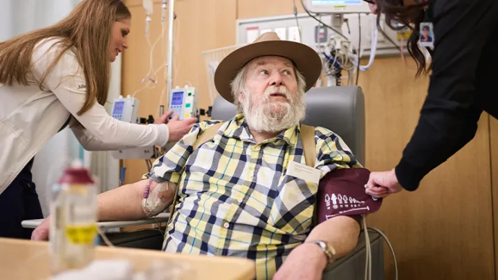
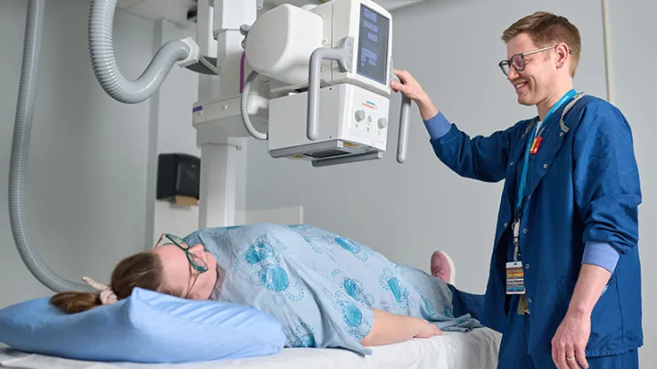

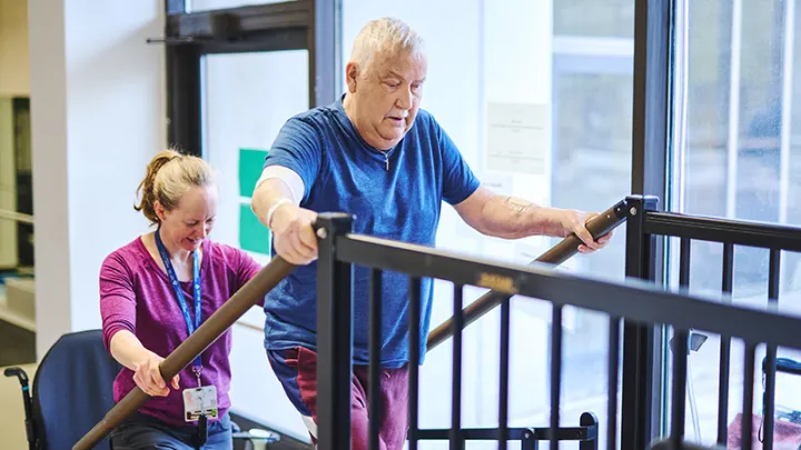

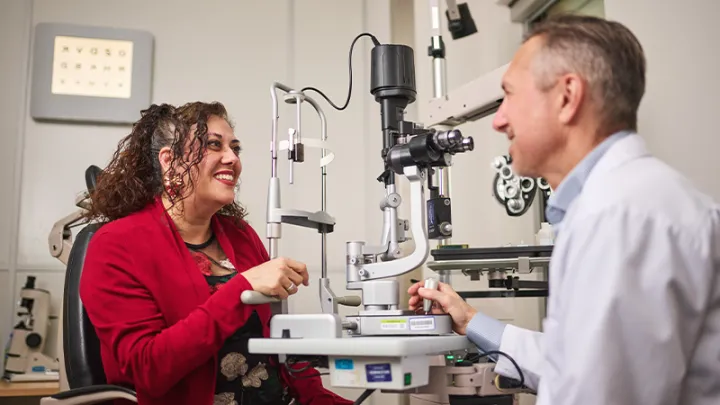

About The Ottawa Hospital
At The Ottawa Hospital, we don’t just serve this community — we belong to this community, and that insight is at the heart of our ambitious plan to reshape the future of health care, reflecting 21st-century needs and innovations, supporting people in the community as well as patients in the hospital.

Get Involved
The Ottawa Hospital strives to be an active part of our community and you can play an integral role in helping make that happen. By donating, volunteering or participating in research, you’re helping The Ottawa Hospital deliver the kind of health care we all want for our loved ones.