

 Print
Print  Share
Share  Text
Text  To reset, hold the Ctrl key, then press 0.
toh
Breast Imaging
Exams Performed at TOH
While screening mammograms are routinely administered to detect breast cancer in women who have no apparent symptoms, diagnostic mammograms are used after suspicious results on a screening mammogram or after some signs of breast cancer alert the physician to check the tissue. Such signs may include:
- A lump
- Breast pain
- Nipple discharge
- Thickening of skin on the breast
- Changes in the size or shape of the breast
A diagnostic mammogram can help determine if these symptoms are indicative of the presence of cancer.
What Are the Benefits and Risks of Screening?
Benefits:
- The benefit of screening is finding cancer early, when it’s easier to treat.
Risks:
- Potential harms from breast cancer screening include pain during the procedure and radiation exposure from the mammogram test itself.
- While the amount of radiation in a mammogram is small, there may be risks with having repeated X-rays.
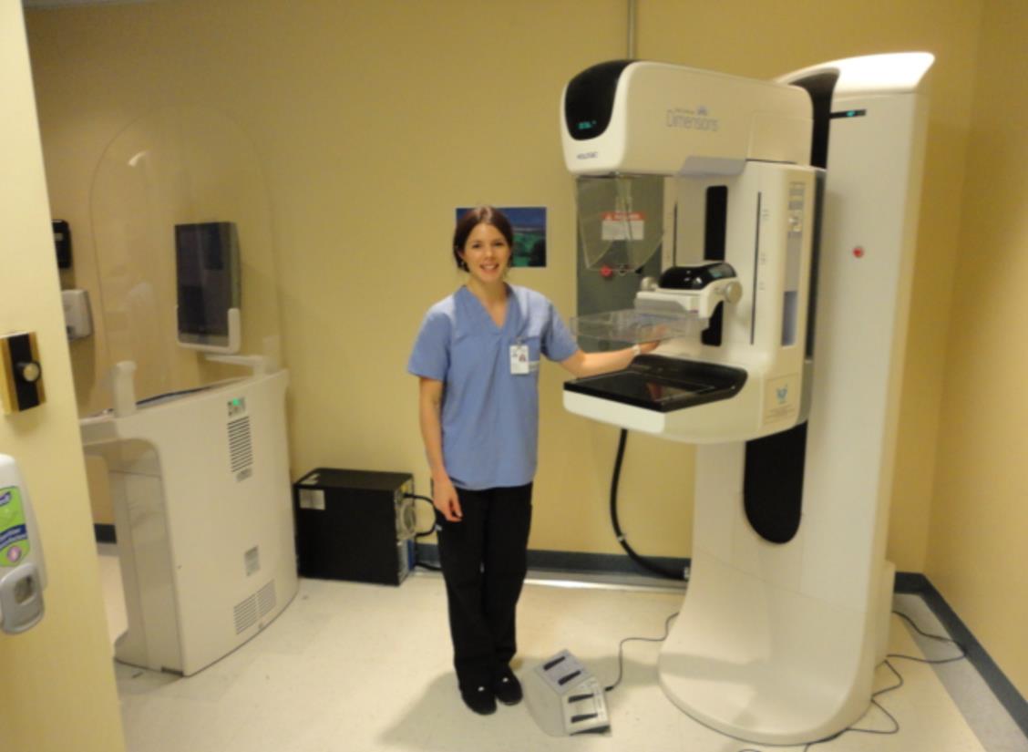
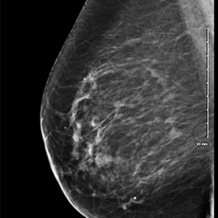
A mammogram is an X-ray picture of the breast. A Mammogram is a simple way for doctors to look for early signs of breast cancer and other health concerns. Regular mammograms are the best way to find breast cancer early, in some cases up to three years before it can be felt.
CC VIEW – VUE CC
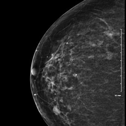
MLO VIEW – VUE OLM
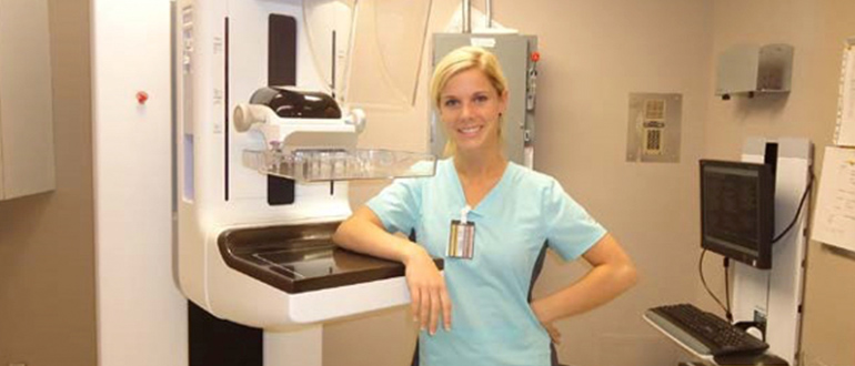
How is a mammogram done?
- Your technologist will ask you to stand in front of a special type of X-ray machine.
- A technologist will then help you to place your breast on a comfortable plastic plate. The technologist will then gently press your breast together with another plate.
- The plates are used to flatten your breast and keep you very still while the x-ray is being taken. Although you will feel some pressure on your breast, the picture will only take a moment to complete.
- You will then be asked to wait a short amount of time while the technologist checks all of your pictures.
- The technologist will then tell you if your X-rays are finished. You will have no side effects from your x-rays.
What does having a mammogram feel like?
- Having a mammogram is a little uncomfortable for most women. The pressure placed on your breasts must be quite firm in order for the x-rays to be very clear.
- It is important to remember, that a mammogram takes only a few moments to complete and the discomfort will soon be over.
- The amount of discomfort you experience depends on the sensitivity of your For most women, this discomfort is not too bad.
- Your breasts may be more sensitive just before or during your period.
- A specially trained doctor, called a radiologist, will read the mammogram. He or she will look at the X-ray for changes in breast tissue.
Safety information
- You should inform technologist if there is any chance that you are pregnant or if you have missed your period recently.
- You should inform the booking staff, before arriving for your appointment, if you have any difficulty standing or require any other form of assistance.
How to Prepare
- Try not to have your mammogram scheduled the week before you have your period or during your period. Your breasts may be more tender or swollen at these times.
- On the day of your mammogram, avoid wearing deodorant, perfume, or These products can affect the quality of your mammogram.
- Since you are required to undress from the waist up for your x-ray pictures, it may be easier if you wear pants and a top instead a dress or skirt.
When will I get my results?
- Once your X-ray images are complete, a radiologist will read your mammogram and report the results to you or your doctor.
- Normally your results will go to your doctor within one week of your mammogram.
- If the radiologist requires any more tests to make your X-rays more clear, you will be contacted by the mammography facility.
- Contact your doctor or the mammography facility if you do not receive your results within 30 days.
What happens if my mammogram is normal?
- Together with your doctor, you may decide how frequently to have Mammograms are an easy and reliable way to maintain good breast health.
- Mammograms work best when they can be compared with previous X-ray. This allows the radiologist to compare them to look for any changes in your breasts.
What happens if my mammogram is abnormal?
- An abnormal mammogram does not always mean that there is cancer.
- It is very common for the radiologist to request a repeat mammogram, an ultrasound or a magnetic resonance procedure to have a more clear picture of your breast tissue.
- You may also be referred to a breast specialist or a surgeon. Again, this does not necessarily mean that you have cancer or need surgery. These doctors are experts in diagnosing breast problems and may request more procedures to help them better understand your breast health.
Who performs the exam?
- A specially trained Medical Radiation Technologist (MRT) will preform your mammogram.
- The MRT and the radiologist work together, to provide you with high quality breast imaging and overall care.
Post exam information
- The radiologist is a specially trained doctor who supervises and interprets radiology examinations. He or she will analyze the images and send a signed report to your doctor who will discuss the results with you. The radiologist may also choose to speak to you in person regarding your results.
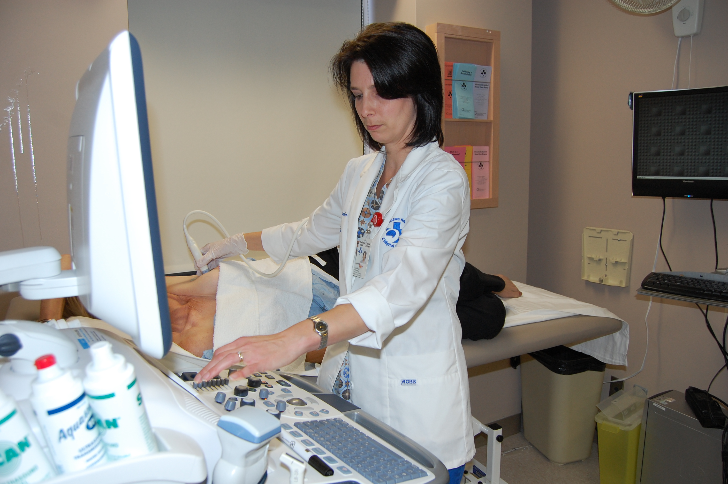
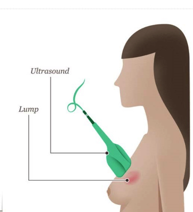
How does an ultrasound help to diagnose a breast lump?
When an unclear area or lump is found in your breast, through a breast self-exam or on a screening mammogram, your doctor may request an ultrasound of the breast tissue. A breast ultrasound is a type of imaging that uses special sound waves to see breast tissue more clearly. Ultrasound imaging does not harm you in any way and cannot be heard by humans. The breast tissue deflects these sound waves and a computer uses this information to create an image of the inside of your breast. Ultrasound is very helpful to the radiologist to know whether a lump is fluid filled or solid.
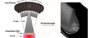
Digital tomosynthesis (pronounced toh-moh-SIN-thah-sis) creates a 3-dimensional picture of the breast using X-rays.
- Digital tomosynthesis is a special test that takes multiple X-ray pictures of each breast from many angles.
- For this test, your breast is positioned in the same way as a mammogram. Only a little pressure is applied to your breast — just enough to keep the breast very still during imaging. The X-ray tube then moves in an arc above your breast while taking numerous pictures.
- The X-ray images are then sent to a computer, where they are put together to produce images that are three dimensional.
- These three dimensional images allows the radiologists to see through layers of tissue and examine unclear areas from many angles.
Benefits can include:
- Earlier detection of small breast cancers that may be hidden during normal mammography
- Greater accuracy in pinpointing size, shape and location of abnormalities
- Fewer unnecessary biopsies or additional tests
- Greater likelihood of detecting multiple breast tumors, which occur in 15% of breast cancer patients
- Clearer images of dense breast tissue
How does a breast MRI help to diagnose breast tissue changes?
During diagnostic breast examinations, it is often helpful for the radiologist to have several different types of studies performed to have a very good understanding of your breast health. If any of your breast imaging procedures are not completely clear, your doctor may recommend a breast MRI (magnetic resonance imaging) to better assess your breast health. During a breast MRI, a technologist will ask you to lie on a special imaging table and then position you into a large tunnel. This tunnel contains a very powerful magnet which is strong enough to create images of your breast tissue. As the magnetic energy passes through your body, you will hear some loud mechanical noises. The technologist will speak to you throughout your images to make sure you are comfortable. The magnetic energy and the noises are not harmful in any way and you will not experience any side-effects.
What is a breast biopsy?
A breast biopsy is a test that removes tissue or sometimes fluid from an area of your breast that your doctor is interested in. The removed cells are examined under a microscope and further tested to check for the presence of breast cancer. A biopsy is the only diagnostic procedure that can definitely determine if a breast area of concern is cancerous. The good news is that 80% of women who have a breast biopsy do not have breast cancer.There are three types of biopsies:
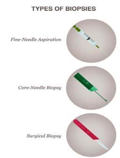
- Fine-needle aspiration
- Core-needle biopsy
- Surgical biopsy
Fine-needle Biopsy
What is fine-needle aspiration?
In most cases, a fine needle aspiration is chosen when the lump is likely to be filled with fluid. If the lump is easy to find or if the doctor suspects that it is a fluid-filled cystic lump, the doctor may choose to do a fine-needle aspiration (FNA). During this procedure, the lump should collapse once the fluid inside has been drawn and discarded. Sometimes, an ultrasound is used to help your doctor guide the needle to the exact site, whereby sound waves create a picture of the inside of the breast. If the lump persists, the surgeon or radiologist, a doctor who specializes in medical imaging such as x-rays and mammograms, will perform a fine needle aspiration biopsy (FNABx), a similar procedure using the needle to obtain cells from the lump for examination.
Core-Needle Biopsy
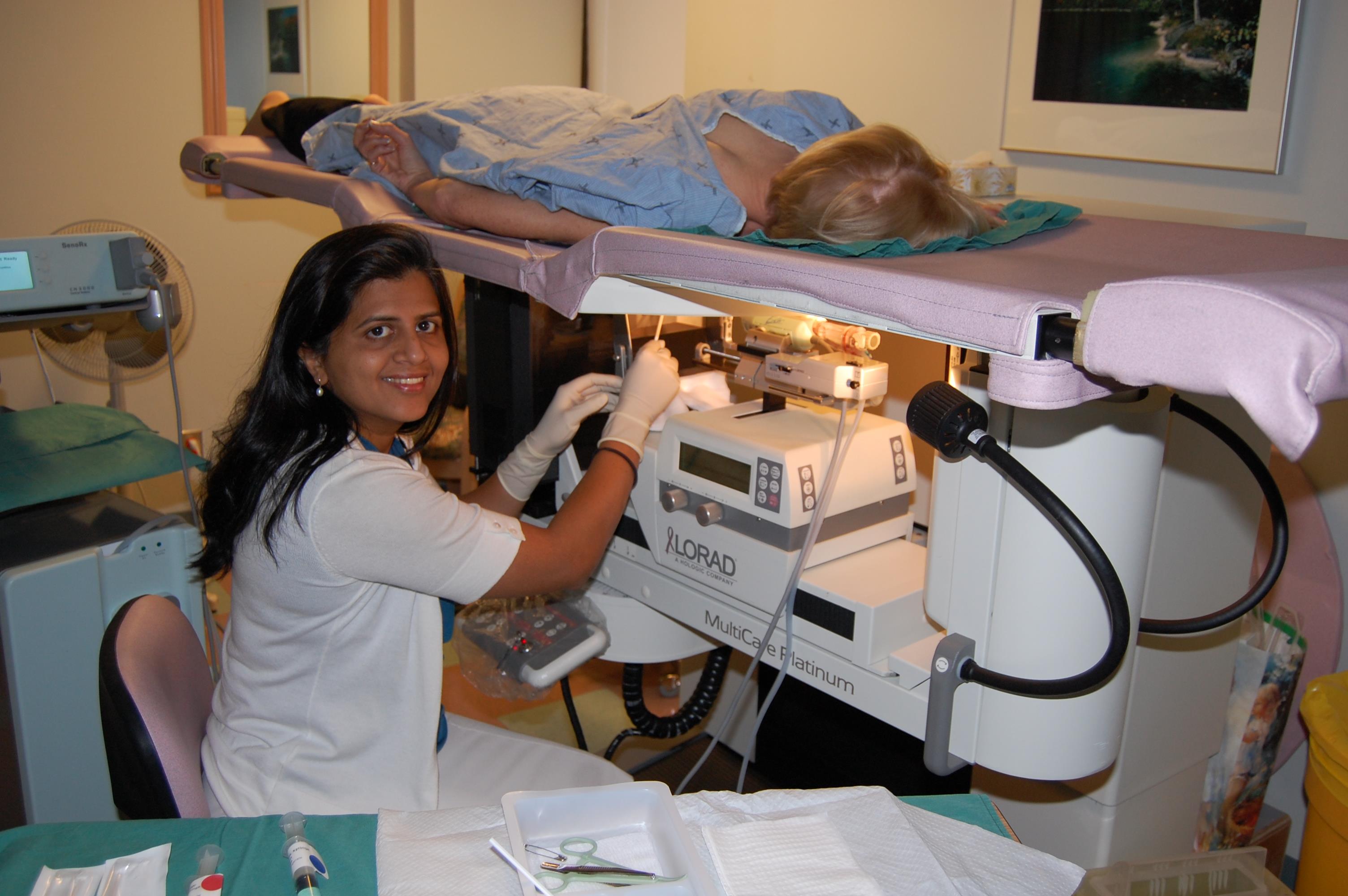
What is a core-needle biopsy?
- Core needle biopsy is the procedure to remove a small amount of tissue from the breast with a larger “core” (meaning “hollow”) needle. To make this procedure more comfortable for you, a small amount of “freezing” or “numbing” medicine is injected into a part of your breast. During the procedure, the doctor may insert a very small marker inside the breast to mark the location of the If surgery is later required, the marker makes it easier for the surgeon to locate the area of concern.
- The radiologist or surgeon performing the core-needle biopsy may use specialized imaging equipment, such as ultrasound, to guide the needle to the desired location.
- If ultrasound is used, the doctor will place an ultrasound probe against your breast to direct the needle. When the doctor chooses to do a stereotactic-guided core-needle biopsy, X-ray equipment and a computer are used to guide the needle. Normally, you will be positioned lying on your stomach on a special table, with an opening for your breast. Sometimes the procedure is done sitting at an upright stereotactic biopsy unit. Your breast will then be compressed in the same way as for a mammogram.
What can be learned from the biopsy results?
- Once the biopsy is complete, a specially trained doctor called a pathologist examines the tissue or fluid samples under a microscope, looking for abnormal or cancerous cells.
- The pathology report, which can take one or two weeks to complete, is then sent to your doctor.
- The report will tell whether the tissue sample has cancer or any other problem. Waiting for your results to return may be challenging, but contains very important information for your doctor.
- Your doctor will review the report with you. If no cancer cells are found, the report will indicate that the cells in the lump are benign, meaning there was no cancer found in your breast.
- However, some type of follow-up or treatment may still be needed, as recommended by the healthcare professional.
- If cancer cells are found, the report will provide more information to help determine the next steps. Your doctor will discuss your treatment choices with you.
- If you need breast cancer surgery, radioactive seed localization (RSL) can help your surgeon to locate and remove your breast tumor, while helping to spare healthy tissue.
- Radioactive seeds help radiologists to pinpoint areas of concern that appear on your mammogram, so your surgeon can remove the abnormal tissue.
- Using ultrasound or mammographic guidance, a radiologist will place a seed into your breast lump.
- The seed is the size of a small grain of rice, and will not be felt by you or your surgeon. The seed is placed in your breast several days before your surgery and will be removed at surgery.
- A type of special imaging is used to see the exact location of the radioactive seed and the abnormal tissue.
- When a radioactive seed cannot be placed, the radiologist may insert a thin wire to mark the area.
What Can I Expect After MY RSL Procedure
- The low radioactivity of the seed is not dangerous to you or others around There are no side effects and you may continue doing everything that you normally do.
- You will have your scheduled surgery up to five days after the RSL procedure.
During the surgery, your surgeon will:
- Use a special device that detects the radioactive “rays” given off by the seed to see the exact location of your tumor.
- Remove your breast tumor and the radioactive seed.
Last updated on: November 10th, 2023